When someone contracts an infection or undergoes a surgical procedure (particularly bone marrow transplantation), doctors track how many T-cells a patient has for immune monitoring.
However, identifying T-cells amongst other biological defenses is a complex process, which is why professionals rely on ID badges to make it a bit easier. And one of the most reliable badges is CD7, found on the surface of the T-cells.
With that said, doctors need accurate procedures and tools to ensure the measurements are accurate. This is why biomedical researchers invest significant resources into developing and testing high-fidelity tools like the CD7 recombinant antibody.
It is understandable that it might be a lot to unpack all at once. This blog will explore the biological significance of CD7 and why the recombinant format is the gold standard for immune monitoring and cancer research.
Understanding CD7 in Detail
CD7 is a member of the immunoglobulin superfamily. It is one of the earliest surface antigens to appear during T-cell ontogeny, showing up even before the T-cell receptor (TCR) is expressed. And this is exactly what makes it an indispensable marker for identifying the very first stages of T-cell development in the thymus.
In addition to being a simple marker, CD7 also plays a functional role in immune costimulation, i.e., it is involved in T-cell and NK-cell activation, helping these cells communicate and respond to threats. Because CD7 is consistently expressed on the vast majority of peripheral blood T-cells, it serves as a “pan-T-cell marker.”
Why the Recombinant Format Matters: Recombinant vs. Hybridoma
For decades, antibodies were produced using hybridoma technology (monoclonal antibodies derived from mouse cells). Yes, these were effective, but these traditional methods often suffer from “batch-to-batch variability”, and in a clinical setting, even a slight variation in antibody affinity can lead to inconsistent immune monitoring results.
To eliminate this inconsistency, recombinant antibodies are preferred. Now, modern companies like AAAbio produce them by cloning the specific antibody genes into high-yield expression vectors (like CHO or HEK293 cells).
In addition to being highly reproducible, these recombinant antibodies do not need animal immunization and are excellent at reducing background noise in complex assays.
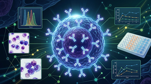
How the CD7 Recombinant Antibody is Applied
This recombinant antibody is used in three ways:
1. Immunohistochemistry (IHC)
In cases of suspected T-cell lymphoma or organ transplant rejection, a blood test isn’t enough; doctors need to see the tissue. For this, a tiny slice of tissue (biopsy) is fixed onto a slide, which is then heated, and then the CD7 recombinant antibody is applied to uncover T-cells. Then, a secondary reagent creates a brown or red stain, allowing a pathologist to see exactly where the T-cells are gathering under a microscope.
2. Flow Cytometry Protocol (Real-Time)
To get a real-time “headcount” of a patient’s immune defenses, labs use flow cytometry to physically count T-cells one by one. First, a blood sample is treated with a CD7 recombinant antibody that has been “tagged” with a glowing dye. After a short incubation, where the antibody latches onto the T-cells, a special liquid is added to clear away red blood cells that might block the view. Finally, the remaining cells are zipped through a high-speed laser; every time a tagged T-cell passes through, it emits a flash of light, allowing a computer to record exactly how many active T-cells are circulating.
3. The ELISA Method: Measuring Soluble CD7
Sometimes, doctors need to measure the specific proteins that T-cells “shed” into the bloodstream, which acts as a smoke signal for immune activation. This is done using the ELISA method, where a “capture” antibody is glued to the bottom of a tiny plastic well to act as a trap. When the patient’s serum is added, any floating CD7 proteins get stuck to the bottom, and a second CD7 recombinant antibody is added to lock them in place from the top—creating a molecular “sandwich.” A final chemical trigger causes the liquid to change color, telling the lab exactly how much protein is present based on the intensity of the shade.